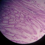
Adenocarcinoma Colon
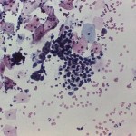
Adenocarcinoma of vagina, depicted in the image below, detected using Papanicolaou stain (Pap stain). Image courtesy of CDC/ A. Elizabeth ... Read More »
Amyloidosis, depicted in the images below, may be identified by the presence of abnormally deposited amyloid proteins in the tissues. ... Read More »
Lipomas, depicted in the images below, may be identified by the presence of cells resembling normal, matured adipocytes with peripheral ... Read More »
Basal cell carcinoma, depicted in the images below, may be identified by the presence of small and large nests of ... Read More »
Squamous cell carcinoma, depicted in the images below, may be identified by the presence of atypical looking squamoid cells having ... Read More »
Leiomyoma, depicted in the images below, may be identified by the presence of whorled bundles of smooth muscle cells having ... Read More »
Thrombosis, depicted in the images below, may be identified by the presence of Lines of Zahn, which show alternate layers ... Read More »
Chronic venous congestion in spleen, depicted in the images below, may be identified by the presence of engorged vessels, some ... Read More »
Chronic venous congestion in liver, depicted in the images below, may be identified by the distention of central vein and ... Read More »


