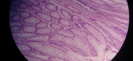
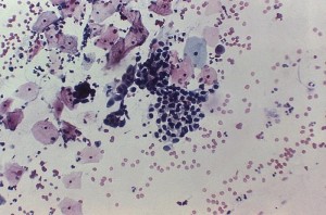
Fatty change, depicted in the images below, can be identified by the presence of small vacuoles containing triglycerides present in cytoplasm around the nucleus of cell. With the increase in severity of disease, these vacuoles merge to form clear spaces that displace the nucleus towards the periphery. Fatty cysts may also be seen.
[smooth=id:69;]


