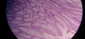
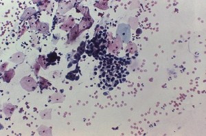
Chronic venous congestion in liver, depicted in the images below, may be identified by the distention of central vein and sinusoids are with RBCs. Some areas of hemorrhage are present as well. Central lobular necrosis due to central vein congestion is seen. Fatty change is due to hypoxia in peripheral hepatocytes.
[smooth=id:84;]
Want a clearer concept, also
Read the article on Chronic Venous Congestion
Compare it with normal histology of liver
See images on Chronic Venous Congestion in Lungs


