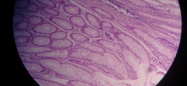
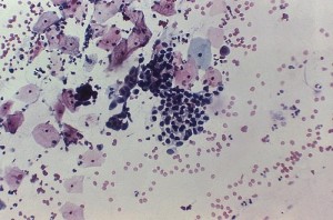
Chronic venous congestion in lungs, depicted in the images below, may be identified by the presence of engorged alveolar capillaries in alveolar septa. There will also be intra-alveolar hemorrhages and alveolar septa and spaces may contain numerous heart failure cells. Alveolar septa undergo fibrosis too.
[smooth=id:83;]
Want a clearer concept, also
Read the article on Chronic Venous Congestion
Compare it with normal histology of lungs


