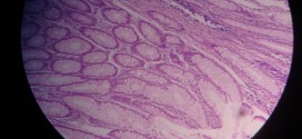
Hydropic change, depicted in the images below, can be identified by tubular cells being swollen and lightly stained. Small, clear vacuoles are present in the cytoplasm, which may be water vacuoles or represent distended endoplasmic reticulum. Due to swelling of cells, lumen of renal tubules becomes narrow or completely obliterated. Swollen tubules compress the microvasculature present between them.
[smooth=id:67;]


