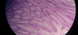
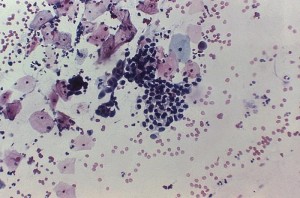
Caseous necrosis, depicted in the images below, can be identified by the presence of necrotic areas, appearing as eosinophilic, amorphous, granular debri composed of necrotic cells. Inflammatory border around the necrotic debri is also seen (granulomatous reaction), which consists of outermost rim of fibroblasts, then T lymphocytes, then macrophages and epitheloid cells and occasional plasma cells.
[smooth=id:71;]Want a clearer concept, also
Read the article on Caseous Necrosis


