Novel Flu H1N1 virus virions, depicted in the image below, are obtained from highly-magnified, digitally-colorized transmission electron micrograph. It is ... Read More »
Novel Flu H1N1 (Swine Flu) Virus
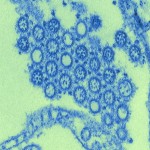
Novel Flu H1N1 virus virions, depicted in the image below, are obtained from highly-magnified, digitally-colorized transmission electron micrograph. It is ... Read More »
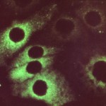
Rubella virus positive serum sample, depicted in the image below, found using fluorescent antibody technique. Rubella virus, responsible for causing ... Read More »
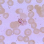
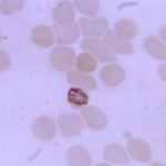
Plasmodium vivax gametocyte, depicted in the image below (1125x), is visible as being reddish in color, and displaying virtually no ... Read More »
Plasmodium vivax merozoites, depicted in the image below, are free from the confines of erythrocyte (red blood cell). Image courtesy ... Read More »

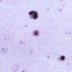
Plasmodium vivax schizont, depicted in the image below, is displaying 14 chromosomal masses. Image courtesy of CDC/ Dr. Mae Melvin ... Read More »
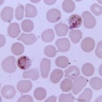
Plasmodium vivax trophozoite, depicted in the image below, is displaying a distinctly pigmented cytoplasm. Image courtesy of CDC/ Dr. Mae ... Read More »
A young, growing Plasmodium malariae trophozoite, depicted in the image below, has just passed its ring-stage in its maturation process. ... Read More »
Plasmodium falciparum immature schizont, depicted in the image below, is obtained on a thin film blood smear micrograph. It is ... Read More »

Bacillus anthracis colonial morphology, depicted in the image below, is found when grown on a medium of chocolate agar, for ... Read More »
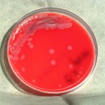
Bacillus anthracis colonial morphology, displayed in the images below, is found when grown on a medium of phenylethyl alcohol agar, ... Read More »


